Plasma membrane: A membrane that allows selective permeability(allowing some substances to pass more easily than others).
Cell membrane: The plasma membrane separates the cell from its surroundings.
Cell membrane mainly contains phospholipids, glycoproteins and cholesterol.
Fluid mosaic model:
"Fluid" because the molecules can move around within their own layer.
"Mosaic" because the protein molecules are dotted within the membrane

W - A phospholipid molecule.
X - A transmembrane protein (can be a channel or carrier protein)
Y - The carbohydrate chain of a glycoprotein.
Z- A cholesterol molecule.
Membrane Proteins
They are of two types,
Intrinsic proteins: Membrane proteins that span fully or partially across the membrane.
Extrinsic proteins: Proteins that are attached on either side of the membrane.
Channel protein
It is an intrinsic protein that aids in facilitated diffusion.
Carrier protein
It is an intrinsic protein that aids in active transport and also facilitated diffusion.
Comparing them:
Similarities:
Both are intrinsic.
Both are specific for their substances.
Both provide pathways for hydrophilic substances.
Differences:
Channel proteins are fixed in shape but carrier proteins can change shape.
Channel proteins only aid in facilitated diffusion but Carrier proteins aid in both active transport and facilitated diffusion.
Functions of membrane proteins
After combining with carbohydrates they form glycoproteins that act as receptors.
Some glycoproteins act as "cell markers" or antigens allowing cell recognition.
Channel proteins and carrier proteins provide pathways for the transportation of polar (hydrophilic substances).
Some proteins inside the cell are attached to a system of protein filaments known as the cytoskeleton.
Membrane Carbohydrate
Many lipid and protein molecules on the outer surface of a cell surface membrane have short carbohydrate chains attached to them forming glycolipids.
Function:
The carbohydrate chains project like antennae into the watery environment surrounding the cell which, by forming hydrogen bonds, stabilizes the membrane structure.
Some glycolipids act as "cell markers" or antigens allowing cell recognition.
They aid in cell-to-cell adhesion.
Cholesterol
A relatively small molecule that has a hydrophobic tail and hydrophilic head.
They fit nearly fit between the phospholipid molecules.
Their heads are at the membrane surface.
Function:
At low-temperature cholesterol increases the fluidity of a membrane.
It does this by preventing the close packing of phospholipid tails.
It prevents the cell from being too rigid and surviving in colder climates.
Cholesterol also prevents the cell from becoming too fluid at a higher temperature.
It does this by interacting with phospholipid tails.
It stabilizes the cells at a higher temperature.
Cholesterol provides mechanical stability to a membrane.
Cholesterol is much less common in plant cells and absent in prokaryotes.
Controlling fluidity of membranes
⦿The more unsaturated the fatty acid is the more fluid the membrane.
This is because the unsaturated fatty acid tails bent and therefore fit together more closely.
⦿The longer the fatty acid tail the less fluid the membrane.
Longer fatty acid tails interact more with each other to fit more closely.
⦿ Higher the temperature the more fluid the membrane is.
Cell signalling
The molecular mechanisms by which cells detect and respond to external stimuli. It includes communication between cells.
The pathway of cell signalling has three main stages:
A stimulus that causes a cell to secrete a specific chemical known as a ligand (e.g. a hormone).
The ligand is transported to the targetted cell by blood.
The ligand binds to the cell surface receptor (glycoproteins) of the target cell.
After the ligand binds with the receptor the following occurs.
The shape of the receptor is changed.

The message is passed into the cell

The message is received by the G-protein

G-protein becomes activated and it activates an enzyme.

The active enzyme breaks down ATP to produce cyclic AMP. This acts as a second messenger.

The second messenger molecule initiates a series of enzyme cascade reactions which amplifies the signal.

Finally, an enzyme is produced that brings the required changes (e.g releasing an enzyme such as protease) within the cell.
For hydrophobic signals, such as steroid hormones, the process is different. During this process, the ligand directly diffuses across the cell membrane and binds to receptors in the cytoplasm or nucleus.
Movement of substances
Diffusion: It is the net movement of particles from a region of their higher concentration to their lower concentration.
The rate of diffusion across cell membrane depends on the following:
The steepness of the concentration gradient
Temperature
Type of molecules or ions
Surface area
Effect of temperature:
The higher the temperature the more kinetic energy molecules have.
The more kinetic energy the faster the rate of diffusion.
Active transport: Movement of substances from a region of lower concentration to their higher concentration against the concentration gradient by using energy from respiration with the aid of carrier proteins.
Osmosis: It is the movement of water molecules from a region of higher concentration to a region of lower concentration, down water potential gradient, through a semi-permeable membrane.
It is a special type of diffusion that only includes water and does not require energy.
Water potential (Ψ): It is the tendency of the water molecules to move.
It is :
Represented by Ψ.
Measured in KPa
Pure water has a potential of 0 KPa.
The more solute is added to water the more negative it becomes.
Hypotonic solution: Solution that has a higher water potential than the specimen in it.
Hypertonic solution: Solution that has a lower water potential than the specimen in it.
Animal cell in a hypotonic solution,
Water from the solution will enter the cell, down the concentration gradient, by osmosis.
The cell starts to expand and as there aren't any cell walls the cell bursts.
Animal cell in a hypertonic solution,
Water from the cell will leave it, down the concentration gradient, by osmosis into the solution.
The cell starts to shrink.
Plant cell in a hypotonic solution,
Water from the solution will enter the cell, down the concentration gradient, by osmosis.
As water enters the vacuole increases in size and pushes the cytoplasm along with the cell membrane against the cell wall.
The cell does not burst due to the presence of a cell wall.
Plant cell in a hypertonic solution,
Water from the cell will leave it, down the concentration gradient, by osmosis into the solution.
As the cell loses water the vacuole decreases in size.
Cytoplasm along with cell membrane shrinks away from the cell wall.
Cell decreases in size and becomes flaccid.
If the cell further loses water the protoplasm (cytoplasm and cell membrane ) loses its shape unable to recover again. This is known as plasmolysis.
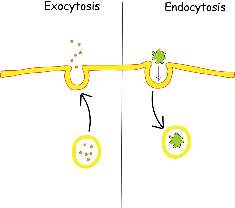
Bulk transport
There are two main types:
Endocytosis
Exocytosis
Process of endocytosis:
Particles (such as pathogens or protein)reach the cell.
It binds to the receptors of the cell membrane that have a complementary shape.
The membrane engulfs the particle by infolding itself.
Vacuole forms that has the particle inside "pinch" off from the cell membrane into the cell.
ATP is used in the process.
Process of exocytosis:
A vesicle is formed around the substance.
The vesicle is formed around the substance.
The vesicle moves towards the cell membrane.
The phospholipid membrane is fluid in nature that allows fusion.
The contents of the vesicle are released.
ATP is used in the process.
⦿During exocytosis, the cell membrane gains some phospholipids.
⦿During endocytosis, the cell membrane loses some phospholipids.
Comments