Cell: Cells are the basic unit of a living organism which can survive on its own.
On the basis of presence or absence of nucleus, cells can be divided into two types:-
Prokaryotic cell: Cells which do not contain any nucleus or any membrane bound organelles.
Example: Bacteria
Eukaryotic cell: Cells which contain a nucleus with a chromosome inside it.
Example: Animal cells, plant cells, fungal cells, protist (plasmodium)
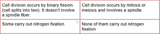
Differences between prokaryotic cells and eukaryotic cells

Organelles: An organelle can be defined as structurally and functionally distinct part of the cell. For example: mitochondria, ribosome, chloroplast, nucleus, centriole.

Cell membrane: Cell membrane is made up of proteins, phospholipids and cholesterol. It is the semi-permeable membrane that encloses the cytoplasm of a cell.
Functions of cell membrane:
It acts as a selectively permeable membrane
It keeps the cell contents together
Allows communication with other cells
Allows recognition of other external substances.
Allows mobility in some organisms e.g. amoebae
* There is no cholesterol in bacterial cell membrane. It is made up of proteins and phospholipids. * Cell membranes can form vesicles.
Nucleus: Nucleus is a double membrane bound organelle, the membrane is known as nuclear envelope.
Nucleus is not continuous and contains gaps with it, known as nuclear pores. The function of nuclear pores is to exchange substances (mRNA, ribosomes, nucleotides) between the nucleus and the cytoplasm.
The densely stained part of the nucleus is known as nucleolus (made up of loops of DNA from several chromosomes). Its function is to manufacture ribosomes using its own DNA.
The jelly like structure in the nucleus is known as nucleoplasm which contains chromosomes in their dispersed form (chromatin).
Nucleus produce RNA to manufacture ribosomes. Chromosomes = DNA + histone protein
Functions of nucleus:
Nucleus is the control center of the cell. It determines metabolism, growth, differentiation, structure and division of the cell.
Nucleus contains the genetic material of the cell in from of chromosomes.
Nucleus acts as the site for transcription.
Mitochondria: Mitochondria is a double membrane bound organelle.
The outer membrane is smooth, inner membrane is folded inwards to form cristae.
Jelly like substance within the mitochondria is known as matrix.
Mitochondria contains smaller circular loop of DNA and 70 S ribosome.
It can change shape and divide and also move about.
They are also flexible and can change shape.
Function of mitochondria:
Mitochondria act as the site for aerobic respiration.
ATP is synthesized in mitochondria.
Chloroplast: Chloroplast is a double membrane bound organelle.
Both membrane are smooth.
Jelly like substance within the chloroplast is known as stroma.
Within stroma there are single membraned structures known as thylakoid membrane; It contains photosynthetic pigment, chlorophyll.
Stock of thylakoid membrane is called grana.
Grana are inter-connected by lamella.
Chloroplasts contain smaller circular loop of DNA and 70 S ribosomes.
Function of chloroplast:
Chloroplast acts as a site for photosynthesis.
Chloroplast contains starch and lipid grains.
Chloroplast synthesizes ATP.
Endoplasmic Reticulum (ER): Endoplasmic reticulum is found near the nucleus and is made up of flattened sacs called cisternae, which are continuous with the nuclear envelope.
The rough endoplasmic reticulum is called so because it has a lot of ribosomes on its outer surface. The RER packages and transports protein that are synthesized in the ribosome.
The RER packages and transports protein synthesized in ribosomes.
The Smooth Endoplasmic Reticulum however do not have ribosomes. SER makes lipids and steroid hormones such as cholesterol and the reproductive hormones estrogen and testosterone.
SER makes lipids and steroid hormones
Lipids: Cholesterol
Steroid hormone: Estrogen and Testosterone
Golgi apparatus: Golgi apparatus is a stack of membrane bound flattened sacs (made up of cisternae).
Golgi apparatus is responsible for the modification, assembly, packaging of proteins received from the ER. These proteins are then transported in vesicles around the cell.
Function of golgi apparatus:
Post translation modification of proteins.
Examples:
Addition of sugar to protein to make molecules known as glycoproteins.
Removal of the first amino acid, methionine, from newly formed proteins to make a functioning protein.
In plants, enzymes in Golgi apparatus converts sugar into cell wall components.
Golgi vesicles are also used to make lysosomes.
Inter relationship between RER and the GA
Proteins are made by the ribosomes on the rough endoplasmic reticulum and are pinched off into vesicles.
Vesicles containing the proteins fuse to the Golgi apparatus where the proteins are modified.
Vesicles containing the modified protein break away from the Golgi apparatus.
Vesicles fuse with the cell surface membrane, releasing the protein outside the cell.
Lysosome: Lysosome is a single membrane bound organelle which is originated from Golgi apparatus.
Lysosomes contain hydrolytic enzymes / digestive enzyme.
Functions of lysosome:
Lysosomes are used to digest bacteria or other microorganisms taken into the cell by phagocytosis or breakdown of unmarked or damaged organelle within the cell.
Lysosomes can digest whole cells as in mammary gland after lactation (breast feeding).
Lysosomes help in replacing cartilage with bones in newborn babies.
The head of the sperm contains a special lysosome called acrosome for digesting the outer membrane of the ovum.
Autolysis
It's when a cell becomes too old to carry out its functions, lysosome gets ruptured releasing hydrolytic enzymes to digest the whole cell.
Ribosome: Ribosome is a non-membrane bound organelle. There are two types of ribosomes, each consists of 2 sub-units:
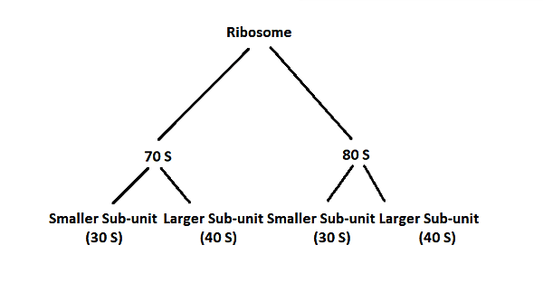
Here, S = Sedimentation (unit of centrifusation)
Each ribosome is made up of rRNA and proteins.
Diameter: 25 micrometers.
Function: Synthesis of protein in the association with RER (rough endoplasmic reticulum)
Eukaryotic cell contains both 80 S and 70 S ribosomes.
Prokaryotic cell contains only 70 S ribosomes.
Eukaryotic cell contains only 80 S within their cytoplasm.
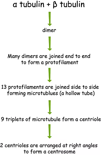
Centriole: Centrioles are non-membrane bound organelles present only in animal cells. They are made up of microtubules which in turn are made up of tubulin protein.
Functions of centriole:
Centrioles act as MTOCs (Microtubule Organizing Centers) for the assembly of microtubules that make up the spindle fibre during nuclear division.
Centrioles found at the base of cilia and flagella are known as basal bodies, act as MTOCs and are responsible for the beating movement of cilia and flagella.
Microtubules: Microtubules are long rigid hollow tubes found in the cytoplasm. They are also very small, 25 micrometres in diameter. (Non-membrane bound cylindrical structure)
Functions of microtubules:
Together with an actin filament and intermediate filament, they make up the cytoskeleton, an essential structural component of cells, which helps to determine cell shape.
Secretory vesicles, organelles and cell components can be moved along the microtubules' outside surface, forming an intracellular transport system e.g. movement of Golgi vesicles during exocytosis.
During nuclear division, a spindle made of microtubules is used to separate chromatids of chromosomes.
Microtubules form part of the structure of the centriole.
The microtubule forms an essential part of the beating movement of cilia and flagella.
Microvilli: Microvilli are tiny, finger-like projections of a cell that increase the cell's surface area for more efficient secretion or absorption.
Functions of microvilli:
Microvilli increase the surface area which is used for-
reabsorption in the proximal convoluted
absorption of digested food into cells lining the gut.
Cilia: They are whip-like beating extensions of many eukaryotic cells. Each cilium is surrounded by an extension of the cell surface membrane.
Structure :
They have two central microtubules and a ring of nine microtubule doublets around the outside.
The two central microtubules are A and B.
The wall of microtubule A is a complete ring with 13 protofilaments.
The wall of microtubule B is an incomplete ring with 10 protofilaments.
Functions of cilia:
Beating mucus upward the throat.
Beating movement in cilia helps the ovum to move in the oviduct.
Cell wall: Formed by running cellulose fibres through a matrix of pectins and hemicellulose. Lignin is also deposited.
Functions of cell wall:
Mechanical strength of cell and plant as a whole.
It prevents a plant cell from bursting when placed in a hypotonic solution.
Forms apoplast (a system of interconnected cell walls) is a major transport route in plants.
Plasmodesmata: They are living connections through neighbouring cell walls.
Functions of plasmodesmata:
Connects neighbouring cell walls.
Allows movement of substances between cells.
Vacuole: Single membrane-bound organelles present only in plant cells.
The membrane is known as tonoplast.
Functions of vacuole:
Storing water.
Storing food.
Storing waste products of cells.
Supporting a cell by maintaining turgor pressure.
Functions of a tonoplast:
Regulating the movement of ions between cytoplasm and vacuole.
Isolating materials that may be harmful to a cell.
Resolution: The ability to distinguish between two objects very close together.
The higher the resolution of an image the greater the detail can be seen.
The resolution of a microscope is half the wavelength of operating radiation.
Higher the wavelength of radiation, the lower the resolution.
The maximum resolution of a light microscope is 200nm.
Points that are closer than 200nm cannot be distinguished by using a light microscope.
The maximum resolution of an electron microscope is 0.5nm.
Transmission vs scanning microscope: A scanning microscope can show the 3D structure of a sample but a transmission microscope shows 2D. Magnification: The number of times greater an image is than the object


Eyepiece graticule: A scale in the eyepiece. It has no unit. Stage micrometre: A scale in the stage. It has a unit of micrometre.
Calibrating a microscope: Given that a specimen measures 6EPG and 10 EPG is equal to 4 SMM. Also, each subunit of SMM is 0.1 mm. Calculate the size of the specimen. As 10 EPG = 6 SMM 1 EPG = 0.4 SMM 6EPG = 0.4 x 6 Converting to millimeters, 0.24 x 0.1 = 0.24 mm = 0.24 x 1000 μm = 240 μm
Bacteria:
Structure :
70S ribosome
Circular DNA
Plasmid
A shorter cytoplasmic extension known as pili that helps bacteria to adhere to the surface.
Virus:
Structure:
Membranous envelope
A protein coat that is known as capsid.
Genetic material which is either DNA or RNA

Comments