Chromosome Structure:
A chromosome is a structural unit that contains genetic information and plays a crucial role in the cell cycle and cell division.
The genetic information within a chromosome is stored in a long, linear molecule of DNA (Deoxyribonucleic Acid).
The DNA in a chromosome is packaged and organized with the help of histone proteins. These proteins help to condense the DNA into a compact structure, making it easier for the cell to manage and utilize genetic information.
During the cell cycle, chromosomes are duplicated in a process called replication. This results in the formation of two identical copies, called sister chromatids, which are held together at a structure called the centromere.
The centromere acts as a central point of attachment between the two sister chromatids, allowing them to be separated during cell division.
To protect the genetic information stored in the DNA, the ends of chromosomes are covered by special structures called telomeres. Telomeres act like caps on the ends of chromosomes, preventing the loss of genetic information during DNA replication and maintaining the stability of the chromosome.

Importance of Mitosis:
Mitosis is important in the production of genetically identical daughter cells, which is crucial for various processes, including:
Growth of multicellular organisms
Replacement of damaged or dead cells
Repair of tissues by cell replacement
Asexual reproduction
During mitosis, the chromosomes are divided evenly between the two daughter cells, ensuring that each cell receives a complete set of genetic information.
This allows for the maintenance of genetic consistency and stability in organisms, which is critical for their survival and growth.
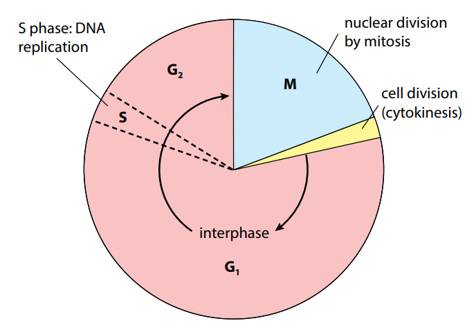
Mitotic Cell Cycle:
The mitotic cell cycle is the process by which a cell divides into two identical daughter cells.
The cell cycle is comprised of several stages, including:
Interphase:
Growth in G1 phase
Growth in G2 phase
DNA replication in S phase
Mitosis:
Prophase
Metaphase
Anaphase
Telophase
Cytokinesis:
Division of the cytoplasm and formation of two distinct cells.
Interphase:
G1 Phase:
This is the first stage of interphase and lasts for several hours to several days, depending on the type of cell.
During this phase, the cell grows and performs metabolic functions.
It also checks for DNA damage, which can occur due to exposure to environmental stressors.
If the DNA is damaged, the cell will halt its progression through the cell cycle and initiate repair mechanisms.
S Phase:
This is the stage of DNA replication, where the chromosomes are replicated to form identical sister chromatids.
During this phase, the DNA is replicated in preparation for cell division.
The entire process takes about 8-12 hours in a typical animal cell.
G2 Phase:
This is the final stage of interphase and lasts for a few hours.
During this phase, the cell grows further and checks for DNA damage.
If the DNA is damaged, the cell will not proceed to mitosis, and instead initiate repair mechanisms.
Mitosis:
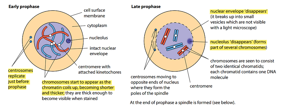
Prophase:
The first stage of mitosis is prophase, which lasts for several hours.
During this stage, the chromosomes condense and become visible, as the histone proteins coil tightly around the DNA.
The spindle fibers, which are microtubules, begin to form and attach to the chromosomes at the centromere.
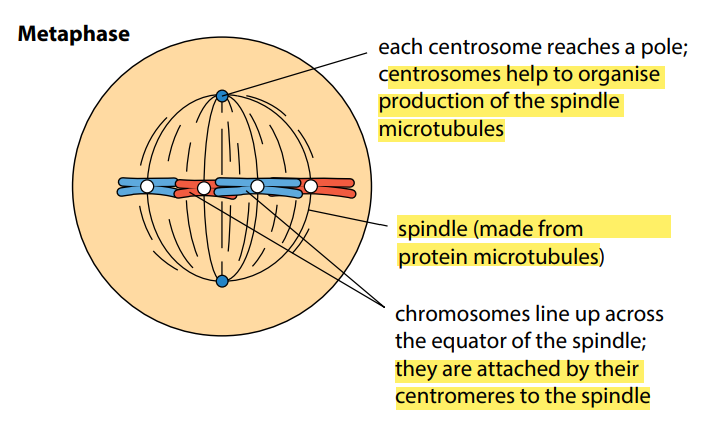
Metaphase:
The next stage is metaphase, which lasts for about an hour.
During this stage, the chromosomes line up at the center of the cell and attach to the spindle fibers.
This is critical to ensure that the chromosomes are separated and distributed evenly into the daughter cells.
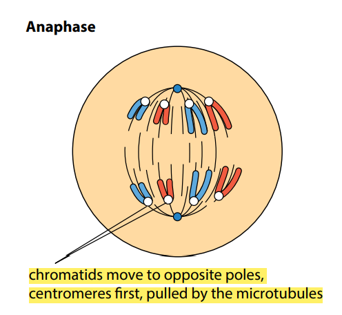
Anaphase:
The following stage is anaphase, which lasts for about an hour.
During this stage, the chromosomes are separated and pulled to opposite poles of the cell by the spindle fibers.
This is a crucial step in the formation of two genetically identical daughter cells.
Telophase:
The final stage of mitosis is telophase, which lasts for a few hours.
During this stage, a new nuclear envelope forms around each set of chromosomes, separating them from each other.
The chromosomes also begin to decondense, as the histone proteins relax their grip on the DNA.
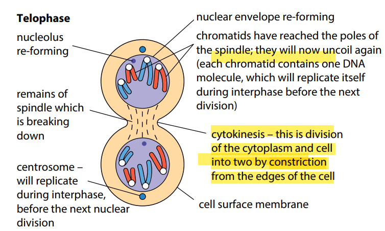
Cytokinesis:
This is the final stage of the mitotic cell cycle and involves the division of the cytoplasm and the formation of two distinct cells.
During this stage, a cell membrane grows between the two sets of chromosomes, forming a division plane.
The cytoplasm then divides along this division plane, separating the two cells.
Role of Telomeres:
Telomeres protect the ends of chromosomes from shortening during DNA replication.
When chromosomes replicate, the DNA replication machinery can't reach the end of the chromosomes.
The telomeres ensure that the ends of the chromosomes are not shortened, and all genetic information is preserved.
Role of Stem Cells:
Stem cells have the ability to differentiate into various cell types and regenerate damaged tissue.
This makes them ideal for use in cell replacement and tissue repair.
When stem cells divide through mitosis, they produce two identical daughter cells, one of which remains a stem cell and the other differentiates into the desired cell type.
This allows for the continuous replacement of damaged or dead cells, promoting tissue repair and maintenance of healthy tissue.
Uncontrolled Cell Division:
Cancer is characterized by uncontrolled cell division and growth of abnormal cells.
Uncontrolled cell division is caused by mutations in genes that regulate the cell cycle, specifically oncogenes.
Oncogenes are normally responsible for promoting cell division and growth. They are involved in regulating the cell cycle and responding to signals that control cell division.
However, mutations in oncogenes can cause them to become overactive, triggering uncontrolled cell division.
As a result, cells continue to divide and grow without normal regulation mechanisms, leading to the formation of a tumour.
Tumours can grow rapidly and can become dangerous if they interfere with normal organ function or spread to other parts of the body.
This is why uncontrolled cell division is a hallmark of cancer and why it is critical to understand and regulate the cell cycle to prevent the development of cancer.
留言